

ADVERTISEMENT
Radial Access for PCI in Children with Obstructive Coronary Artery Disease
ABSTRACT: Percutaneous coronary intervention (PCI) is rarely performed in the pediatric population for obstructive coronary disease occurring secondary to non-atherosclerotic conditions. We report two cases of children treated with PCI utilizing radial access. Both cases employed a strategy of deep conscious sedation, left radial access, and minimal use of cineangiography. Radial access in children undergoing PCI can be performed safely, effectively, and comfortably with minimal radiation exposure and may offer a safety advantage over femoral access in this smaller patient population.
VASCULAR DISEASE MANAGEMENT 2012:9(9):E159-E162
Key words: pediatric cardiac catheterization, percutaneous coronary intervention, radial artery access
____________________________________________________________
Percutaneous coronary intervention (PCI) is occasionally necessary in the pediatric population to treat obstructive coronary artery disease due to Kawasaki’s Disease, external compression associated with a variety of congenital conditions, and coronary allograft vasculopathy (CAV).1 We report two cases of PCI in children utilizing radial access.
Case Reports
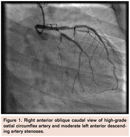 Case 1. A 14-year-old female with history of orthotopic heart transplantation 7 years prior for primary dilated cardiomyopathy was referred for PCI from Children’s Hospital Boston. Routine post-transplant angiography 1-week prior to PCI revealed progressive CAV with a newly noted high-grade proximal left circumflex stenosis (Figure 1). She had been complaining of chest heaviness and shortness of breath with moderate exertion. The patient weighed 105 lbs (47.7 kg) and her height was 62 inches (158 cm). She had prior cardiac catheterizations via femoral access without major complications. Her medications included aspirin, diltiazem, enalapril, furosemide, spironolactone, pravastatin, tacrolimus, and mycophenolate. She received a loading dose of clopidogrel 300 mg the day before her planned PCI. The modified Allen’s test utilizing plethysmography of the left radial artery was normal (Grade A).
 Prior to the sterile prep for the procedure, the pelvis was wrapped in a lead apron and deep conscious sedation was obtained with propofol administered by cardiac anesthesiology. A 5 Fr x 11 cm hydrophilic sheath was inserted into the left radial artery, 2.5 mg of intra-arterial verapamil was administered, and the patient was systemically anticoagulated with heparin. The left main coronary artery was intubated with a 5 Fr EBU 3.0 mm guide catheter (Medtronic). Under fluoroscopic guidance the left circumflex was wired with a 180 cm x 0.014-inch middleweight coronary guidewire, pre-dilated with a 2.5 mm compliant balloon, and ultimately treated with a 3.0 mm Cypher drug-eluting stent (Cordis). Post-dilation was performed with a 3.25 mm non-compliant balloon. Angiography confirmed an excellent final result (Figure 2).
The sheath was removed immediately post-PCI and hemostasis was obtained with compression using a radial compression device. Careful attention was given to ensure patent hemostasis of the radial artery after compression device placement as described by Pancholy et al.2 A total radiation dose of 449 mGy was required for completion of the procedure. She tolerated the procedure well and was discharged home the next day. At 1-week follow-up she was free of symptoms with a normal neurovascular examination at the access site and hand. Her left radial artery was patent by physical exam. Overall, she was pleased with radial access given that her prior femoral procedures had been associated with moderate discomfort.
 Case Report 2. A 16-year-old female patient with history of orthotopic heart transplantation 10 years prior for ventricular septal defect and secondary dilated cardiomyopathy was referred for PCI from Children’s Hospital Boston. Routine post-transplant angiography revealed rapidly progressive transplant vasculopathy compared to an angiogram performed 6 months prior. She had developed a significant stenosis in the mid left anterior descending artery (LAD) (Figure 3) as well as a significant stenosis in the circumflex artery. She was recently diagnosed with post-transplant lymphoproliferative disorder and therefore was not a candidate for repeat transplantation. She had no specific cardiovascular symptoms but had been experiencing progressive fatigue.
 The patient weighed 103 lbs (47 kg) and her height was 58 inches (147 cm). She underwent prior cardiac catheterizations via femoral access without major complications. However, her previous procedures were associated with significant discomfort and anxiety requiring general anesthesia. Her medications included aspirin, enalapril, pravastatin, tacrolimus, and mycophenolate. She received a loading dose of clopidogrel 300 mg the day before her planned PCI. The Allen’s test with modified plethysmography of the left radial artery was normal (Grade A).
 Prior to the sterile prep, the pelvis was wrapped in a lead apron and deep conscious sedation was obtained with propofol administered by cardiac anesthesiology. We obtained arterial access with a micro-puncture system and the patient was given 2.5 mg of intra-arterial verapamil via a 4 Fr x 5 cm dilator. She was systemically anticoagulated with heparin. We attempted to use a sheathless system by advancing a 5 Fr EBU 3.0 mm guide catheter utilizing a 4 Fr x 125 cm MP diagnostic catheter as an introducer. The whole system was advanced over a 0.035-inch guide wire that had been previously advanced to the ascending aorta. However, there was significant resistance in advancing the catheter through the proximal radial artery. Ultimately a 5 Fr x 11 cm hydrophilic sheath was inserted into the left radial artery. The guide catheter could then easily advance into the ascending aorta, allowing for intubation of the left main coronary artery. The LAD was wired with a 0.014-inch x 180 cm middleweight coronary guidewire. The lesion was pre-dilated with a 2.0 mm compliant balloon and ultimately treated with a 2.5 mm Cypher drug-eluting stent. The stent was post-dilated with a 2.5 mm non-compliant balloon resulting in an excellent angiographic result. Next, the circumflex artery was wired and directly stented with a 2.25 mm Cypher drug-eluting stent. Cineangiography confirmed an excellent result (Figures 4 and 5). The sheath was removed and the access site was compressed with a radial compression device with care given to ensuring patent hemostasis. A total radiation dose of 358 mGy was required for the procedure. On the following day, however, she had tenderness over the left radial artery access site and a pulse was difficult to palpate. The reverse Allen’s test at the bedside suggested that the radial artery might have occluded. However, at 1-week follow-up, her pain had significantly improved and a reverse Allen’s test showed patency of the radial artery. Pseudoaneurysm was not present based on physical examination.
Despite the temporary discomfort she experienced at her access site, she felt that radial access was more comfortable with easier recovery compared to her prior femoral diagnostic procedures.
Discussion
The recently published RIVAL study showed that radial access was associated with low rates of bleeding complications and reduced overall vascular complications compared to femoral access in adults undergoing PCI for acute coronary syndromes.3 Additionally, there was a strong patient preference for future procedures to be performed via the radial approach in patients who underwent both radial and femoral access. The trial also showed that radial access cases were associated with longer fluoroscopic times compared to femoral access.
We believe that the benefits of radial access for PCI may apply to the pediatric population. Risks of vascular complications for transfemoral access are highest among patients with the lowest and highest body mass indices (BMI), whereas moderately obese patients (BMI 30-36) have the lowest risk.4 This “J” shaped relationship of BMI and complication rates suggests that the pediatric population may be at higher risk for bleeding complications associated with PCI given their smaller BMI.
When considering radial access in the pediatric population, there are several procedural considerations that may improve the success and safety of the procedure. First, deep sedation administered by anesthesia (or consideration of general anesthesia) should be strongly considered. Children may be more prone to anxiety with procedures in general, and a deeper than standard level of sedation will eliminate anxiety and hopefully reduce the likelihood of spasm in these smaller radial arteries.
Consideration should be given to utilizing the left radial artery rather than the right radial artery in order to facilitate catheter engagement. Because the ascending aorta will be shorter in the pediatric population compared to the adult population, there may be more difficulty in engaging standard guiding catheters when approaching from the right arm. The catheters will have to take a “retroflexed” bend when exiting the innominate artery into the ascending aorta. When working with a shorter ascending aorta, manipulation of catheters to intubate the left main coronary artery can be more challenging. When approaching the ascending aorta from the left arm, the catheter will take a more femoral-like route to the ascending aorta and will likely advance into the left cusp with less manipulation. In a non-randomized observational study of adults undergoing diagnostic catheterization, Kawashima et al showed that procedural times were shorter for left radial access versus right radial access.5 This was further suggested in the TALENT trial, which showed that left radial access may be associated with less fluoroscopic time compared to right radial access during diagnostic coronary angiography.6
In-lab vascular ultrasound should be used for obtaining access when physical examination suggests a small radial artery. With direct visualization of the artery and the access needle, the operator can be certain of the puncture site being in the center of the artery and grazing of the artery can be minimized. This may help reduce the incidence of radial artery spasm.
Utilizing the smallest access catheter possible to complete the procedure is imperative. The majority of PCI can be completed through a 5 Fr guiding catheter advanced within a 5 Fr sheath. However, the arteriotomy size can be reduced further utilizing a sheathless approach.7 For example, a 5 Fr guide is used as the sole catheter entering the wrist and advanced to the coronary artery. The outer diameter of a 5 Fr guide is just greater than 3 Fr and by minimizing the catheter to artery ratio, the risk of radial artery occlusion can be reduced. The sheathless approach is somewhat limited in that 5 Fr hydrophilic guiding catheters and specialized dilators are not available in the United States. Dedicated hydrophilic guides designed specifically for sheathless access may improve success rates for transradial PCI in the pediatric population.
Finally, minimization of radiation exposure is vitally important in this age group given their young age and the likelihood for future cineangiographic procedures. Shielding of the pelvis should be standard and can be easily accomplished since the femoral access areas will not need to remain sterile. Minimization of cineangiographic imaging can be accomplished with liberal storing of fluoroscopic images, a functionality that is now available on most newer generation angiographic imaging equipment.
Radial access for pediatric interventions has been reported in a case series of 12 children and adolescents after heart transplant (mean age 16.7 years, mean weight 55.2 kg).8 In this series using 5 Fr sheaths, the procedure could not be completed due to severe radial artery spasm in 3 children despite their older age and higher weight. This was likely due to reliance on local anesthesia alone in 70% of the cases. This difference underlines the importance of adequate sedation where cannulation of a small radial artery is required. Adequacy of sedation and caliber of the radial artery based on palpation were more important than patient age in our decision to proceed with radial puncture. In select cases, ultrasound of the radial artery would be helpful in making this decision.
In summary, radial access for PCI is safe and feasible and may offer a reduction in vascular complications and improved patient comfort compared to femoral access in the pediatric population. Even with the strategies mentioned above, radial access may not be possible in all pediatric patients. The patients presented in this review were older and not far from small adult size. Radial access for coronary angiography and PCI may be challenging in much younger and smaller patients and therefore, patient selection will be important in ensuring successful radial PCI in the pediatric population.
References
- Shaddy RE, Revenaugh JA, Orsmond GS, Tani LY. Coronary interventional procedures in pediatric heart transplant recipients with cardiac allograft vasculopathy. Am J Cardiol. 2000;85(11):1370-1372.
- Pancholy S, Coppola J, Patel T, Roke-Thomas M. Prevention of radial artery occlusion-patent hemostasis evaluation trial (PROPHET study): a randomized comparison of traditional versus patency documented hemostasis after transradial catheterization. Catheter Cardiovasc Interv. 2008;72(3):335-340.
- Jolly SS, Yusuf S, Cairns J, et al; for the RIVAL trial group. Radial versus femoral access for coronary angiography and intervention in patients with acute coronary syndromes (RIVAL): a randomised, parallel group, multicentre trial. Lancet. 2011;377(9775):1409-1420.
- Cox N, Resnic FS, Popma JJ, Simon DI, Eisenhauer AC, Rogers C. Comparison of the risk of vascular complications associated with femoral and radial access coronary catheterization procedures in obese versus nonobese patients. Am J Cardiol. 2004;94(9):1174-1177.
- Kawashima O, Endoh N, Terashima M, et al. Effectiveness of right or left radial approach for coronary angiography. Catheter Cardiovasc Interv. 2004;61(3):333-337.
- Sciahbasi A, Romagnoli E, Burzotta F, et al. Transradial approach (left vs right) and procedural times during percutaneous coronary procedures: TALENT study. Am Heart J. 2011;161(1):172-179.
- Youn YJ, Yoon J, Han SW, et al. Feasibility of transradial coronary intervention using a sheathless guiding catheter in patients with small radial artery. Korean Circ J. 2011;41(3):143-148. Epub 2011 Mar 31.
- Irving C, Zaman A, Kirk R. Transradial coronary angiography in children and adolescents. Pediatr Cardiol. 2009;30(8):1089-1093.
____________________________________________________________
From the Brigham and Women’s Hospital, Boston, Massachusetts.
Disclosure: The authors have completed and returned the ICMJE Form for Disclosure of Potential Conflicts of Interest. Dr. Shah reports that he has received honoraria from Boston Scientific. Dr. Winston reports no conflicts of interest regarding the content herein.
Manuscript received January 13, 2012, provisional acceptance given February 16, 2012, final version accepted March 21, 2012.
Address for correspondence: Pinak B. Shah, MD, Cardiovascular Division, Brigham and Women’s Hospital, 75 Francis Street, Boston, MA 02115, USA. Email: pbshah@partners.org
